By Richard Gunderman, MD PhD
When many people hear the word apocalypse, they picture four remorseless horsemen bringing death and destruction during the world’s final days. In fact, the Greeks who introduced the word over 2,000 years ago had no intention of invoking the end times. Instead the word apocalypse, which is composed of the roots for “away” and “cover,” means to pull the cover away, to reveal, and to see hidden things. The idea is not merely that we can bring things shrouded in darkness to light, or make visible what was once invisible. These interpretations imply no prior effort to conceal. With apocalypse, there is a clear connotation that what is unseen has been intentionally hidden.
The contemporary medical field of radiology exhibits both revelatory and apocalyptic features. Anyone who has seen x-ray, ultrasound, CT, or MR images of the human body knows that we now routinely peer inside it without cutting it open. Hundreds of thousands of patients each year, who would once have undergone diagnostic surgeries intended to determine what ails them, can now be evaluated in a non-invasive fashion. For example, it is now possible completely to assess the extent of abdominal trauma patients’ injuries with a CT scan, only operating on the small number whose injuries are associated with severe, ongoing blood loss or the interruption of blood flow to a vital organ.
This ability offers huge benefits to the patient and savings to the healthcare system. Important risks, costs, and downstream effects of surgery can be completely avoided. The patient whose imaging findings don’t indicate surgical therapy and doesn’t undergo an operation is spared risks of anesthesia, infection, and bleeding. While CT scans are expensive compared to doing nothing, they constitute a small fraction of the combined cost of anesthesiologist’s and surgeon’s fees, operating room and recovery room time, and prolonged hospitalization and post-operative recovery. And the patient does not go through life with a surgical scar or increased risk of developing a bowel obstruction.
No one who knows anything about medicine would doubt for a moment that the advent of radiology has completely transformed the way physicians care for patients. In fact, a poll conducted at the turn of the millennium revealed that the two most important medical innovations in the latter half of the 20th century were the introduction of CT and MRI scanning. Nothing was introduced into medicine during that time period without which it would be more difficult to imagine providing top-notch care to patients. Fields such as neurology, neurosurgery, emergency medicine, trauma surgery, and oncology would be almost unrecognizable without such technologies.
What can x-rays, ultrasound, CT, and MRI scanners reveal? The answer is simple yet profound — every one of the most important categories of human disease. Infectious and inflammatory disorders generally demonstrate swelling and increased contrast enhancement, appearing brighter than normal tissues. Traumatic injuries usually appear as loss of blood flow to tissues, associated in some cases with hemorrhage. The same can be said for vascular disorders, such as heart attack or stroke, where the blood flow to vital tissues is interrupted. And cancers generally appear as masses that displace or replace normal tissues in the lung, colon, breast, prostate gland, and so on.
The contributions of medical imaging can be regarded as a form of revelation, illuminating the otherwise invisible inner structure and function of the human body. The only alternative ways to visualize such structures is to insert an endoscope, which makes parts of the body such as the linings of the respiratory and digestive tracts directly visible, or to use a scalpel, which necessarily entails damage to normal tissues. In both cases, some form of anesthesia is generally needed, and there are risks of perforation, bleeding, and infection. Of course, the radiation associated with CT scans entails a tiny theoretical risk of cancer, but MRI and ultrasound involve essentially no health risk.
Does it make sense to consider radiology not merely revelatory but also apocalyptic? In other words, would we be justified in saying that the interior of the human body is not only invisible but actually hidden? There are a number of grounds for answering these questions in the affirmative. One obvious sense concerns the fate of a pre-19th century ancestor whose internal anatomy had become accessible to the eye. Whether a trauma victim or a surgical patient, any person whose brain, heart, or intestines had seen the light of day would likely be dead or dying, and those who weren’t would suffer a very high risk of death from infection over the ensuing days (Figure).

From a pre-historical perspective, the exposure of such internal anatomy would necessitate so much destruction of normal tissues, such a large blood loss, and so high a probability of infection that it would be essentially incompatible with life. So far as we know, it was only relatively recently in human history, really only in the last few centuries, that physicians and scientists had gained sufficient knowledge of the structure and function of the human body, as well as the existence of pathogens such as bacteria, to be able to prevent and treat problems such as internal blood loss and bacterial infection. Only by keeping their insides hidden could human beings survive.
Consider also the milieu intérieur, a term coined in the 19th century by the great physiologist Claude Bernard. It refers to the body’s remarkable ability to maintain a relatively stable internal environment in the face of huge swings in external conditions. Summer or winter, gorging or fasting, awake or asleep, we maintain a remarkably consistent internal temperature, blood pH, and glucose concentration, and so on. Bernard and his followers believed that the skin, the linings of our digestive and respiratory tracts, and the immune system play a vital role in maintaining the internal conditions of life. To disrupt such boundaries, for example by a severe burn, is to put the organism at grave risk.
Both nature and man labor to keep the hidden hidden. If the insides are brought into direct contact with the outside, the organism dies. Confronted by a knife-wielding assailant, our impulse to self-preservation provides ample evidence of the depth of the instinct to protect our interior. And this is especially true of our most vital constituents. We naturally make use of less vital parts, such as the arms and legs, to protect the most essential ones, such as our head, neck, and torso. When such inner parts do become directly visible, many people experience a deep, even visceral sense of revulsion, some even becoming nauseous, dizzy, or passing out.
Radiology, then, represents more than a form of revelation. It is apocalyptic, an uncovering of the hidden. As such, it walks a kind of tightrope. On the one hand, it transgresses some of our most deeply seated natural boundaries, revealing what eons of natural selection and millennia of cultural evolution have established as a sanctum sanctorum, off limits to the eyes. Yet it does so in a most remarkable way, making it possible to transgress such boundaries without breaking them down, and for one of the noblest of purposes, to diagnose, stage, treat, and monitor the recurrence of disease. Above all, such transgressions involve not life’s taking but its protection and restoration.
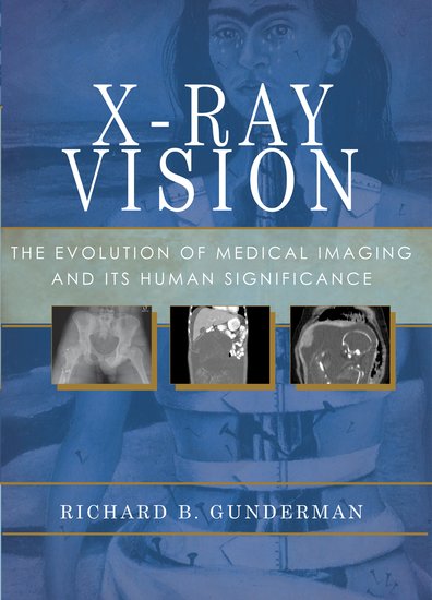
Richard Gunderman, MD PhD, is a Professor of Radiology, Pediatrics, Medical Education, Philosophy, Liberal Arts, and Philanthrophy at Indiana University, Indianapolis, Indiana, and winner of the 2012 Alpha Omega Alpha Robert Glaser Distinguished Teacher Award. He is also the author of X-Ray Vision: The Evolution of Medical Imaging and Its Human Implications.
Subscribe to the OUPblog via email or RSS.
Subscribe to only science and medicine articles on the OUPblog via email or RSS.


Brother, have you seen? The ‘fix’ of the SGR [HR 4015] requires all imaging requests to be electronically pre-approved through HHS. All. Starting in 2015. In 2018, the top 5% of “a users” called indiscriminate order ers will be fined. Read it.